Why advanced imaging matters
Accurate diagnosis is the foundation of accurate treatment. Conventional 2D dental X-rays show teeth and bone in a single plane — but the human jaw is three-dimensional. Structures like nerve canals, root anatomy, sinus proximity and bone density are invisible in 2D, yet critical to safe implant placement, root canal treatment, and surgical planning.
CBCT (cone beam computed tomography) gives us a complete 3D volumetric view of your jaw, teeth, sinuses and surrounding structures. At Balaji's Best Dental, this technology is in-house. You don't need to visit a separate radiology centre, wait days for results, or pay for separate facility fees. Your scan is done at the clinic and reviewed immediately.
CBCT vs OPG — which is used when?
OPG — Orthopantomogram
A panoramic 2D X-ray of the entire upper and lower jaw, all teeth, and surrounding bone in a single image. Fast, low-dose and ideal for a full-mouth overview.
CBCT — Cone Beam CT
A full 3D volumetric scan of the jaw. Shows bone thickness, nerve location, root morphology and sinus anatomy in three dimensions — essential for complex cases.
Implant planning
CBCT is used for all implant cases — to measure available bone precisely, locate the inferior alveolar nerve, and virtually plan implant position before surgery.
Root canal assessment
CBCT reveals extra canals, root curvatures and periapical infections with far greater detail than standard X-rays — critical for complex or retreatment cases.
Orthodontic planning
OPG provides an overview of all erupting and impacted teeth. CBCT is added for impacted canines, skeletal assessment and airway evaluation.
Oral surgery & TMJ
CBCT is used for wisdom tooth proximity to the nerve, jaw cysts, TMJ (jaw joint) assessment and pre-surgical planning by our maxillofacial surgeon.
The imaging process
No preparation required
No fasting, no injections, no contrast agents. Simply remove metal jewellery from your head and neck area before the scan.
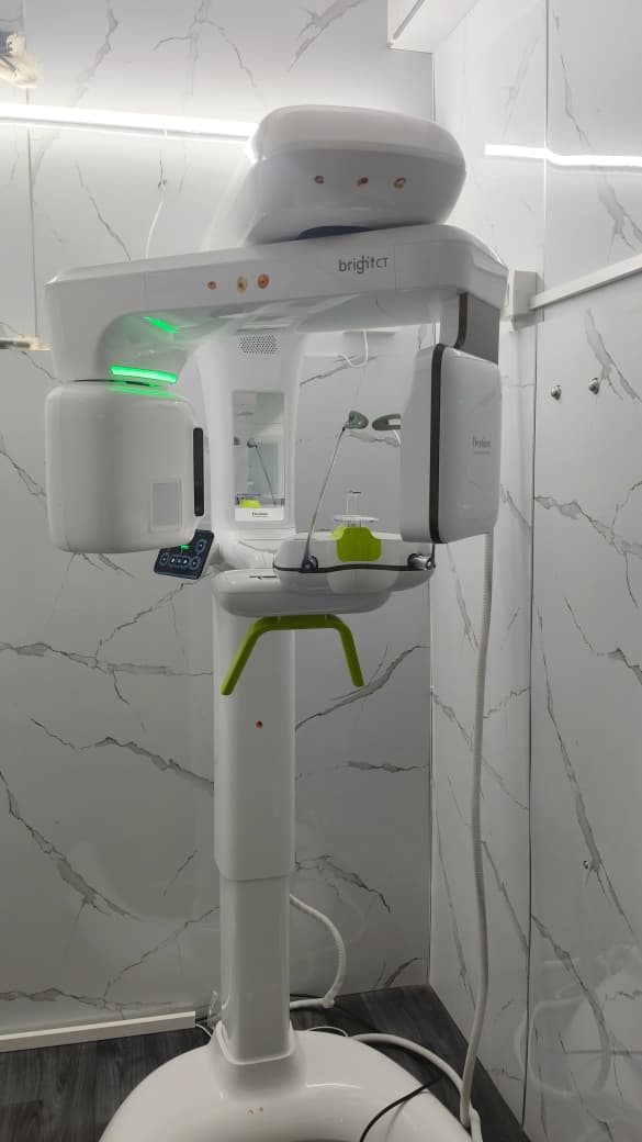
Positioning
You stand or sit inside the imaging unit. Chin and forehead rests guide you into position. The entire process is comfortable and takes under 2 minutes.
Scan acquisition
The rotating arm completes its pass in under 30 seconds. You stay still; the machine moves around you. There is no enclosed space and no discomfort.
Image reconstruction
The scan data is processed into a full 3D model viewable from any angle. DICOM files are available for second opinions or specialist referrals.
Clinical review & treatment planning
Your clinician reviews the images with you — explaining what they show, how they inform your treatment, and what the recommended plan is.
When do you need a CBCT or OPG?
- Before dental implant placement — to assess bone volume, density and nerve position
- Complex or retreatment root canals — to visualise extra canals and infection extent
- Impacted wisdom teeth — to confirm proximity to the inferior alveolar nerve
- Impacted canines and orthodontic planning
- Jaw pain or TMJ (jaw joint) assessment
- Jaw cysts, tumours or pathology evaluation
- Pre-surgical planning for oral and maxillofacial procedures
- General full-mouth review — especially as a new patient baseline
Why choose Balaji's Best Dental?
In-house — no referral delay
CBCT and OPG done at our clinic. No trips to a separate radiology centre. Results are available immediately and reviewed with your treating clinician the same day.
Used for every implant case
Dr. Balaji uses CBCT for every implant case — not just selected ones. Sub-millimetre planning accuracy is what enables our consistent implant outcomes across 4,200+ cases.
DICOM files provided
Your scan data is yours. We provide DICOM files on request for second opinions or referrals to specialists elsewhere.
Low-dose protocol
Our scanner uses optimised low-dose protocols appropriate to the clinical question — the smallest field of view and lowest dose that meets diagnostic needs.